Clinical Summary
A 43-year-old woman presents with complaints of longstanding, occasionally bloody, diarrhea. Colonoscopy shows innumerable mucosal polyps, which are biopsied. Due to the patient’s symptoms and colonoscopic findings, the decision is made to perform a total colectomy. The gross specimen shows diffuse mucosal erythema and numerous polyps, without evident disease elsewhere.
Master List of Diagnoses
- Cap polyposis
- Crohn disease with pseudopolyposis
- Drug-induced colitis with pseudopolyposis
- Familial adenomatous polyposis
- Ulcerative colitis with pseudopolyposis
Archive Case and Diagnosis
This case first appeared as Performance Improvement Program in Surgical Pathology (PIP) 2020, Case 28, and is ulcerative colitis with pseudopolyposis in the colon.
The information provided in this case was accurate and correct at the time of publication in 2020. Any changes in terminology since the time of publication may not be reflected in this case.
Criteria for Diagnosis and Comments
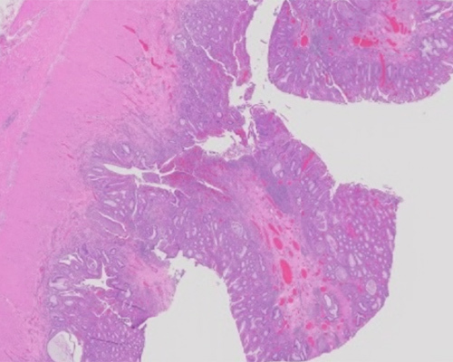
Sections show a segment of colon with numerous polyps that demonstrate marked inflammation and injury, with crypt architectural distortion, crypt abscesses, and expanded lamina propria. The inflammation extends into the submucosa and very focally into the muscularis propria and beyond. For the most part, however, the disease is relegated to the mucosa and submucosa. Small granulomas, when present, appear to represent a response to ruptured crypts. Overall, the findings are most consistent with ulcerative colitis with extensive pseudopolyposis.
Ulcerative colitis (UC) is one of two forms of inflammatory bowel disease, the other being Crohn disease (CD, aka regional enteritis). The disease is more common in Caucasians, with roughly equal prevalence in men and women. Incidence estimates are as high as 20 cases per 100,000 persons. UC has a bimodal distribution, with peak onsets at approximately 15 - 30 years of age and 50 - 70 years of age. However, patients may present in the “gap” between the peaks, including with longstanding disease not previously brought to medical attention (as in this patient). Symptoms lapse and remit; they include abdominal pain and bloody, mucoid diarrhea.
UC can involve most or all of the large intestine, including the rectum, involving the entire mucosa in the affected length (ie, no “skip lesions”). Mucosal changes indicative of chronicity include lamina propria expansion by an increased lymphoplasmacytic infiltrate, crypt distortion, crypt branching, crypt dropout, crypt foreshortening, basal plasmacytosis, and Paneth cell metaplasia in the distal colon. In active disease, acute inflammation is superimposed upon chronic changes; it may be mild (cryptitis), moderate (crypt abscesses), or severe (mucosal ulceration). In most cases, UC only damages the mucosa, with the rest of the colon unaffected. In patients with severe UC (such as this case), there may be a hint of minor mural inflammation.
Treatment in UC begins with medications aiming to keep the inflammation in check, including anti-inflammatory medications such as sulfasalazine and mesalamine. Immunosuppressant medications are needed for some patients, such as azathioprine and infliximab. Surgery is reserved for severe, medically refractory cases.
Crohn disease has a similar epidemiology to UC and may also cause inflammatory pseudopolyposis. The main differences between UC and CD are that the latter can affect the entire gastrointestinal tract and can affect areas discontinuously (“skip lesions”), and the inflammation can involve the entire wall. Microscopically, CD therefore demonstrates transmural chronic inflammation, often forming prominent lymphoid aggregates. Other findings include aphthous ulcers and epithelioid granulomas away from areas of crypt rupture. Since none of these findings are present in the current case (aside from occasional mild mural inflammation due to disease severity), the findings are more consistent with UC than CD.
Cap polyposis is a rare form of injury-induced colonic polyposis. The inflammatory polyps all bear a characteristic “cap” of granulation tissue at their surface. While little is known about this condition, the symptoms are similar to UC, and limited tissue sampling via colonoscopy might suggest UC with pseudopolyposis as a possible diagnosis. However, the polyps in the current case clearly do not have caps of granulation tissue.
Drug-induced colitis can mimic inflammatory bowel disease clinically and microscopically, requiring careful clinicopathologic correlation. Many drugs can cause colitis mimicking inflammatory bowel disease, including nonsteroidal anti-inflammatory drugs and mycophenolate mofetil. This diagnosis has become more common with the advent of checkpoint inhibitors, which can prolong the life of patients with metastatic malignancies but have several side effects. These include CTLA4 inhibitors (ipilimumab) and PD1 inhibitors (nivolumab, pembrolizumab). Colitis can occur any time during (or even after) treatment. Their patterns of histologic injury can vary, but the presence of increased intraepithelial lymphocytes or increased crypt apoptosis should suggest the possibility of drug-induced injury. To date, colonic pseudopolyposis has not been described in this setting.
Familial adenomatous polyposis (FAP) is one of several genetic syndromes that lead to development of premalignant polyps. Patients harbor an APC gene mutation and develop hundreds or thousands of colonic tubular adenomas. While this case would mimic FAP on colonoscopy, the lack of dysplasia in any of the polyps rules out FAP.
Supplementary Questions
- Which of the following is more common in ulcerative colitis than Crohn disease?
- Aphthous ulcers
- Inflammatory pseudopolyposis
- Mucosa-limited inflammation
- Mucosal granulomas
- Skip lesions
- Which of the following may be an indication for surgery in ulcerative colitis?
- Concomitant primary sclerosing cholangitis
- Crypt rupture granulomas
- Medically refractory disease
- Polypoid low-grade dysplasia
- Rectal involvement
- Colitis may occur secondary to inhibitory antibodies targeting what protein?
- APC
- CTLA2
- HER2
- PD1
- TNF-alpha
References
- Cerilli LA, Greenson JK. The differential diagnosis of colitis in endoscopic biopsy specimens: a review article. Arch Pathol Lab Med. 2012;136(8):854-864.
- Gonzalez RS, Salaria SN, Bohannon CD, Huber AR, Feely MM, Shi C. PD-1 inhibitor gastroenterocolitis: case series and appraisal of 'immunomodulatory gastroenterocolitis'. Histopathology. 2017;70(4):558-567.
- Kini GP, Murray I, Champion-Young J, et al. Cap polyposis mistaken for Crohn's disease: case report and review of literature. J Crohns Colitis. 2013;7(3):e108-111.
- Rosty C. The Role of the Surgical Pathologist in the Diagnosis of Gastrointestinal Polyposis Syndromes. Adv Anat Pathol. 2018;25(1):1-13.
- Tontini GE, Vecchi M, Pastorelli L, Neurath MF, Neumann H. Differential diagnosis in inflammatory bowel disease colitis: state of the art and future perspectives. World J Gastroenterol. 2015;21(1):21-46.
Answer Key
- Mucosa-limited inflammation (c)
- Medically refractory disease (c)
- PD1 (d)

