Introduction
As a biomarker, HER2 (human epidermal growth factor receptor 2), encoded by ERBB2, has evolved from a poor prognostic indicator in breast cancer to a significant precision oncology target. As a 185-kDa EGFR family tyrosine kinase lacking an endogenous ligand, HER2 dimerizes, especially with HER3, to activate PI3K-AKT-mTOR and MAPK-ERK signaling pathways. HER2 dysregulation occurs through ERBB2 amplification with protein overexpression, most commonly in breast and gastric cancers, or through activating kinase domain mutations, particularly in non–small cell lung cancer (NSCLC). This biology underpins current diagnostic and therapeutic approaches.
A major milestone came in April 2024, when the FDA granted accelerated approval to fam-trastuzumab deruxtecan-nxki (T-DXd) for any unresectable or metastatic HER2-positive (IHC 3+) solid tumor, representing the first tumor-agnostic approval based on protein expression, expanding a category previously limited to genomic biomarkers like MSI-H and NTRK fusions.1 With this shift, HER2 testing, traditionally utilized in breast and gastric pathology, has now expanded to encompass a broad array of solid tumors. This expansion places pathologists at the forefront of accurate assay interpretation and the integration of morphologic, molecular, and therapeutic considerations across varied clinical settings. This article highlights the growing therapeutic landscape and reviews fundamental principles of HER2 testing, with a focus on immunohistochemistry (IHC) scoring.
The HER2-Directed Treatment Landscape
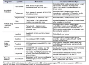
Since the approval of trastuzumab in 1998, the HER2-targeted therapeutic landscape has evolved substantially. Four drug classes now comprise FDA-approved therapies: monoclonal antibodies, antibody–drug conjugates (ADCs), small-molecule tyrosine kinase inhibitors (TKIs), and bispecific antibodies (Table 1). The tumor-agnostic approval of T-DXd for any HER2-positive (IHC 3+) solid tumor constitutes a landmark development, extending tissue agnostic, biomarker-driven therapy beyond genomic alterations to include HER2 protein overexpression.1
Table 1. FDA-Approved HER2-Targeted Therapies by Drug Class
HER2/ERBB2 Alterations and Testing Modalities
HER2 dysregulation arises through three principal mechanisms: gene amplification, protein overexpression, and activating mutations, each with distinct tumor-type distribution and therapeutic implications.
Gene Amplification and Protein Overexpression
ERBB2 gene amplification leads to increased copy number and HER2 protein overexpression on the cell membrane. This mechanism predominates in breast cancer (~15%-20%) and gastric/GEJ adenocarcinoma (~12%-24%)2. Pan-cancer sequencing studies reveal ERBB2 amplification in approximately 3%-5% of all solid tumors, with enrichment in bile duct, bladder, and gynecologic malignancies.1,3-4
Activating Mutations
Activating ERBB2 mutations arise independently of gene amplification and have particular clinical significance in NSCLC, where they occur in 1%-4% of cases.5,6 These alterations involve multiple regions of the receptor, most commonly the tyrosine kinase domain and extracellular domains. Notably, ERBB2-mutant tumors infrequently exhibit ERBB2 amplification or HER2 overexpression, highlighting the critical role of comprehensive next-generation sequencing (NGS) in identifying affected patients.
Testing Modalities
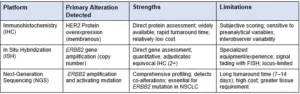
ERBB2/HER2 assessment integrates three complementary platforms (Table 2). Immunohistochemistry (IHC) remains the cornerstone of assessment, providing direct protein evaluation at the cell membrane. In situ hybridization (ISH), including fluorescence (FISH) and chromogenic (CISH) methods, assesses ERBB2 gene amplification and is routinely used to resolve equivocal (IHC 2+) results. NGS enables detection of ERBB2 amplification and activating mutations and is particularly critical in NSCLC.5,6 Selection of the appropriate diagnostic assay depends on the underlying molecular alteration and tumor type.
Table 2. HER2/ERBB2 Testing Modalities: Strengths and Limitations
HER2 IHC Scoring Criteria
Since its initial utilization in breast cancer diagnostics, HER2 IHC scoring has undergone progressive refinement. The original ASCO-CAP guidelines, first introduced in 2007 and revised in 2013, 2018, and 2023, defined HER2 positivity in breast carcinoma as strong, complete circumferential membrane staining (3+) in greater than 10% of invasive tumor cells, a criterion reflecting the uniform membranous expression characteristic of mammary ductal epithelium.7,8 The subsequent expansion of HER2-targeted therapy to gastric and gastroesophageal junction (GEJ) adenocarcinomas, following the ToGA trial,2 prompted the development of the Hofmann scoring system (2008) and later CAP-ASCP-ASCO guidelines (2016), which established modified criteria that accept basolateral or lateral (U-shaped) membranous staining patterns as positive to accommodate architectural complexity and biologic heterogeneity of gastrointestinal tumors.9,10
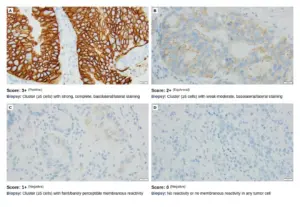
This distinction is clinically significant: applying breast-specific circumferential criteria to gastric cancer would result in underscoring and potential exclusion of patients who may benefit from HER2-targeted therapy. The 2024 FDA approval of T-DXd for any HER2-positive (IHC 3+) solid tumor has further expanded this diagnostic paradigm, requiring pathologists to apply tumor-appropriate scoring algorithms across all solid tumors. This evolution underscores the need for pathologists to be fluent in multiple scoring systems and to explicitly document which criteria were applied in each case. Table 3 provides a comprehensive comparison of currently available HER2 IHC scoring criteria across solid tumors. Figure 1 demonstrates HER2 IHC scoring in gastroesophageal adenocarcinoma.
Table 3. Comprehensive Comparison of HER2 IHC Scoring Criteria Across Solid Tumors
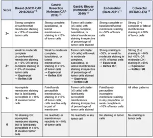
Figure 1. HER2 IHC Scoring in Gastroesophageal Adenocarcinoma
Challenges and Future Directions
The expansion of HER2-targeted therapies across tumor types has brought renewed attention to interpretive challenges in HER2 IHC scoring. Intratumoral heterogeneity, preanalytical variables including fixation time and tissue processing, and interobserver variability in assessing staining intensity and membrane patterns remain ongoing quality assurance considerations.10 These factors underscore the importance of rigorous laboratory standardization, adherence to validated preanalytic parameters, and consistent application of established scoring protocols.
For malignancies beyond breast, gastric, colorectal, and endometrial carcinomas, validated organ-specific IHC scoring criteria are not yet available. In current practice, gastric resection scoring criteria, which accept basolateral and lateral membrane staining patterns, have been adopted for other solid tumors, consistent with the methodology employed in the pivotal DESTINY-PanTumor02 trial.1 While this approach provides a practical framework for contemporary clinical decision-making, continued refinement and prospective validation of tumor-specific scoring algorithms will be important as experience with pan-tumor HER2 testing expands.
Within this evolving landscape, the pathologist occupies a central role not only in technical assay interpretation, but also in integrating the morphologic context, selecting the appropriate scoring algorithm, and determining therapeutic eligibility. Ensuring standardized, high-quality HER2 testing across solid tumor types is essential to achieve accurate biomarker classification and equitable patient access, recognizing that organ-specific criteria remain unavailable for many malignancies.
Key Takeaways
- HER2 testing has expanded from organ-specific to tumor-agnostic indications, placing new interpretive demands on pathologists.
- HER2 IHC scoring criteria are tumor-specific and not interchangeable; inappropriate application can lead to patient misclassification.
- For tumors lacking validated scoring system, gastric resection criteria currently provide a pragmatic interim framework
- Pathologists play a central role in assay interpretation, algorithm selection, and documentation, directly impacting therapeutic eligibility.
- Ongoing practice refinement and prospective validation are needed as pan-tumor HER2 testing continues to evolve.
⚑ CLINICAL PEARL: HER2 Scoring for Pan-Tumor Indications
Pan-Tumor T-DXd Eligibility
The FDA tumor-agnostic approval for trastuzumab deruxtecan (T-DXd; April 2024) is based on HER2 IHC 3+ status only. Unlike breast and gastric cancers, reflex ISH is not required to determine eligibility for this pan-tumor HER2-directed therapy.
Scoring Criteria for Other Solid Tumors
Validated organ-specific HER2 IHC scoring guidelines do not currently exist for biliary tract, bladder, ovarian, or other non-breast/gastric/colorectal/endometrial malignancies. Since the DESTINY trials applied gastric resection scoring criteria, current consensus practice is to adopt these criteria (accepting basolateral/lateral membrane staining patterns) when evaluating HER2 status in other solid tumors. This approach should be documented in the pathology report.
Guideline Update
CAP gastric HER2 IHC scoring guidelines are currently under review; however, no organ-specific IHC scoring criteria have been validated for many tumor types at this time. Readers are encouraged to consult their institutional policies and the latest publicly available guidance for updates.
References
1. Meric-Bernstam F, Makker V, Oaknin A, et al. Efficacy and safety of trastuzumab deruxtecan in patients with HER2-expressing solid tumors: primary results from the DESTINY-PanTumor02 phase II trial. J Clin Oncol. 2024;42(1):47-58.
2. Bang YJ, Van Cutsem E, Feyereislova A, et al. Trastuzumab in combination with chemotherapy versus chemotherapy alone for treatment of HER2-positive advanced gastric or gastro-oesophageal junction cancer (ToGA): a phase 3, open-label, randomised controlled trial. Lancet. 2010;376(9742):687-697.
3. Ismail A, Jani CT, Jahan N, et al. HER2 alterations across solid tumors: implications for comprehensive testing. Oncologist. 2025;30(9):oyaf258.
4. Harding JJ, Fan J, Oh DY, et al. Zanidatamab for HER2-amplified, unresectable, locally advanced or metastatic biliary tract cancer (HERIZON-BTC-01): a multicentre, single-arm, phase 2b study. Lancet Oncol. 2023 Jul;24(7):772-782.
5. Li BT, Smit EF, Goto Y, et al. Trastuzumab deruxtecan in HER2-mutant non-small-cell lung cancer. N Engl J Med. 2022;386(3):241-251.
6. Le X, Kim T, Loong H, et al. Sevabertinib in Advanced HER2-Mutant Non-Small-Cell Lung Cancer. N Engl J Med. 2025;393:1819-1832.
7. Wolff AC, Hammond MEH, Allison KH, et al. Human epidermal growth factor receptor 2 testing in breast cancer: American Society of Clinical Oncology/College of American Pathologists clinical practice guideline focused update. J Clin Oncol. 2018;36(20):2105-2122.
8. Wolff AC, Somerfield MR, Dowsett M, et al. Human epidermal growth factor receptor 2 testing in breast cancer: ASCO-College of American Pathologists guideline update. J Clin Oncol. 2023;41(22):3867-3872.
9. Hofmann M, Stoss O, Shi D, et al. Assessment of a HER2 scoring system for gastric cancer: results from a validation study. Histopathology. 2008;52(7):797-805.
10. Bartley AN, Washington MK, Colasacco C, et al. HER2 testing and clinical decision making in gastroesophageal adenocarcinoma: guideline from the College of American Pathologists, American Society for Clinical Pathology, and American Society of Clinical Oncology. Arch Pathol Lab Med. 2016;140(12):1345-1363.
11. Buza N. HER2 testing in endometrial serous carcinoma: time for standardized pathology practice to meet the clinical demand. Arch Pathol Lab Med. 2021;145(6):687-691.
12. Valtorta E, Martino C, Sartore-Bianchi A, et al. Assessment of a HER2 scoring system for colorectal cancer: results from a validation study. Mod Pathol. 2015;28(11):1481-1491.