This program offers at least two challenging cases each month from the CAP Performance Improvement Program in Surgical Pathology. For each case, you will use a virtual microscope whole slide image of a surgical specimen to diagnose diseases.
You will be presented with information on each case including: specimen source, clinical history, laboratory findings, and a representative whole slide image of the surgical specimen. After you complete each case, you will receive a case summary with three multiple-choice questions that relate to the case.
Please note: DigitalScope® is supported with Microsoft Internet Explorer 11.0 (limited support for IE 9 and 10) or later, or FireFox 4.0 or later, or Safari 3 or the latest Google Chrome version.
 Posterior Chest Wall
 Posterior Chest Wall
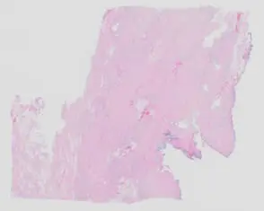
A 59-year-old woman presents with a newly identified back mass. She denies any tenderness or recent trauma. A firm, immobile mass is palpated in the deep soft tissue at the inferior angle of the left scapula. Overlying skin changes are absent. Intraoperatively, the mass is noted to be firm and indistinct, extending from the scapula to the underlying rib.