
This case was originally published in 2021. The information provided in this case was accurate and correct at the time of initial program release. Any changes in terminology since the time of initial publication may not be reflected in this case.
Clinical History
The patient is a 16-year-old boy presenting with two-month history of lower back and leg pain. Spine MRI revealed an intradural, extramedullary mass at the level of the cauda equina at L4. The mass measured 2.8 cm in greatest dimension and showed homogenous intermediate signal on T1-weighted images, heterogenous signal on T2-weighted images, and diffuse enhancement post-contrast (Image A).
Tissue Site
Spinal cord, cauda equina, L4
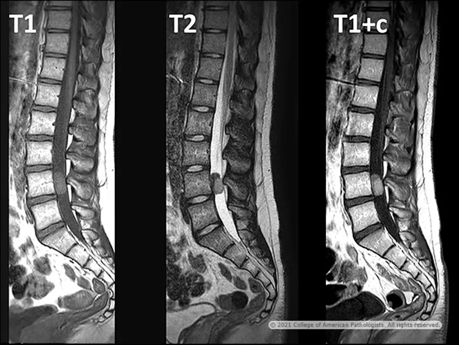
Image A: T1-weighted, T2-weighted, and T1-weighted post-contrast MRI.
Whole Slide Image
The whole slide image provided is an H&E-stained section from cauda equina tumor resection.
Questions
- Which diagnosis is most likely?
- Clear cell meningioma
- Hemangioblastoma
- Myxopapillary ependymoma
- Schwannoma
- If the patient in this case has a germline pathogenic variant, it most likely involves which gene?
- TP53
- SMARCA4
- SMARCB1
- SMARCE1
- Which of the following is most likely to show strong diffuse immunoreactivity for inhibin?
- Clear cell meningioma
- Hemangioblastoma
- Myxopapillary ependymoma
- Schwannoma
Discussion and Diagnosis
Meningiomas are common tumors of the CNS and have a wide variety of histological appearances. Clear cell meningioma (CCM) is a rare variant that accounts for only 0.2% of all meningiomas. CCMs tend to affect children and young adults, and they commonly occur in the cerebellopontine angle and spinal cord, particularly at the level of the cauda equina. CCMs are designated as World Health Organization (WHO) grade 2 tumors, as they tend to show aggressive behavior with high rates of recurrence and a propensity to disseminate through the CSF.
CCMs display MRI features similar to other meningiomas; they are generally isointense on T1-weighted images and iso- or hyperintense on T2-weighted images, with strong homogenous or heterogenous contrast enhancement (Image A). While most meningiomas are adherent to the dura, several cases of nondural-based CCMs have been reported, and therefore they may lack a dural tail sign on MRI.
Histologically, CCMs often display patternless or sheeting architecture (Image B). Tumor cells are round to polygonal with clear, glycogen-rich cytoplasm (Image C). Whorls are uncommon and if present, tend to be poorly formed, and psammoma bodies are frequently absent. Perivascular and interstitial collagen is frequently present and can form small concentric collagenous knots. Collagen may also coalesce into large acellular zones of hyalinization (Image D), which in some cases may predominate over the neoplastic cells. Brightly eosinophilic amianthoid-like collagen may also be present. Extracellular deposits of petaloid tyrosine-rich crystals are an uncommon feature (Image E and Image F). Like other subtypes of meningiomas, the clear cell variant is immunoreactive for EMA (Image G) and somatostatin receptor 2A (SSTR2A), may show nuclear staining with progesterone receptor, and is immunonegative for GFAP (Image H) and S100 (Image I). Glycogen within tumor cell cytoplasm can be highlighted with PAS positivity (Image J) that is diastase sensitive.

Image B: H&E. |
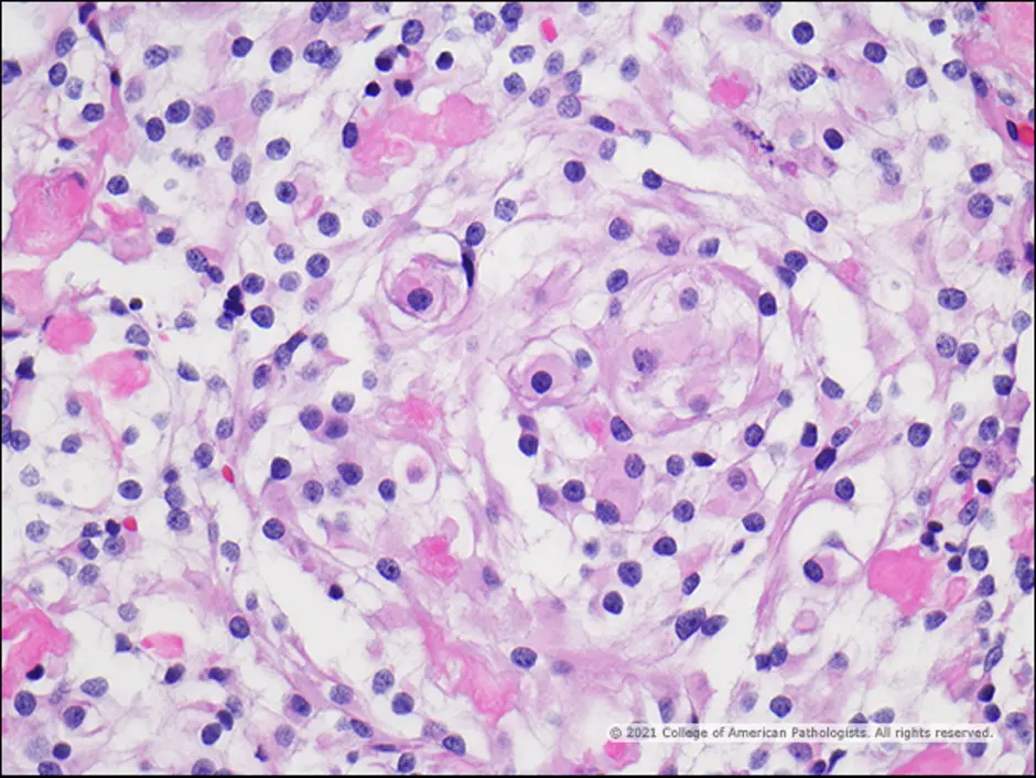
Image C: H&E. |
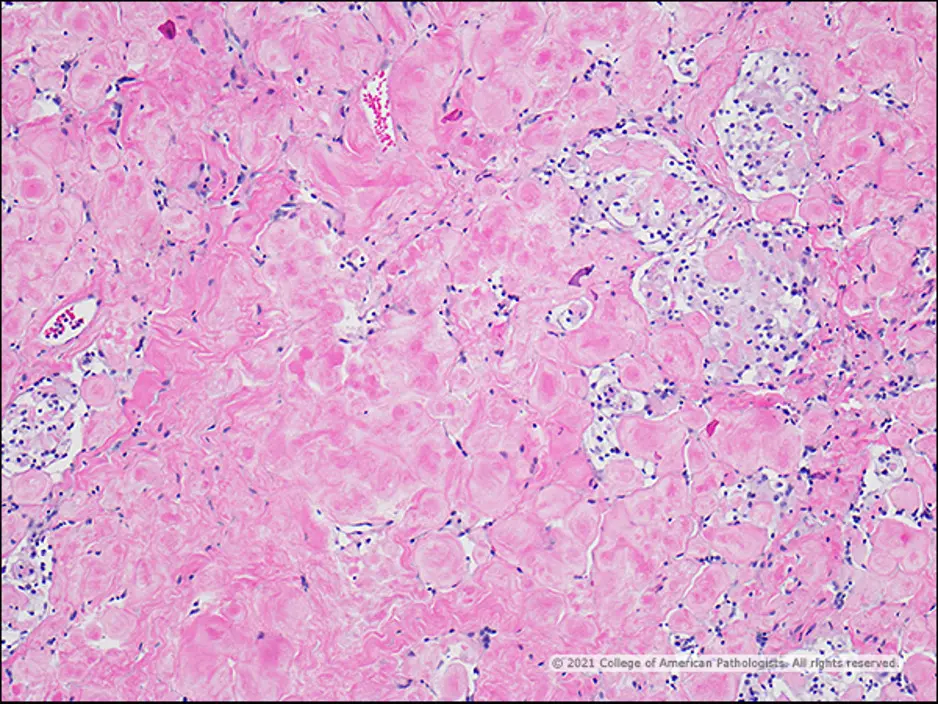
Image D: H&E. |
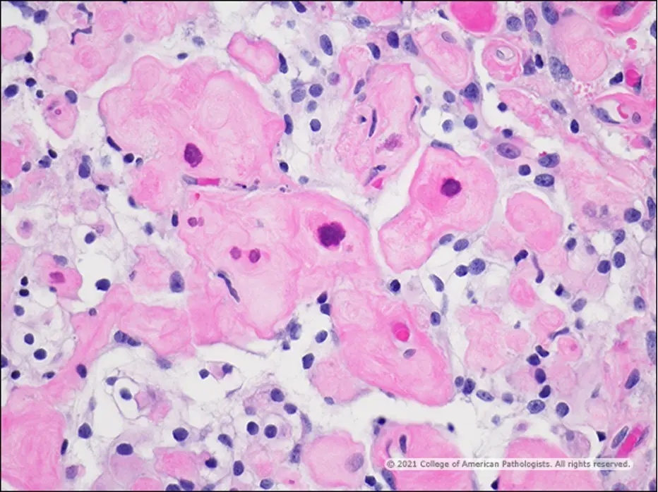
Image E: H&E. |
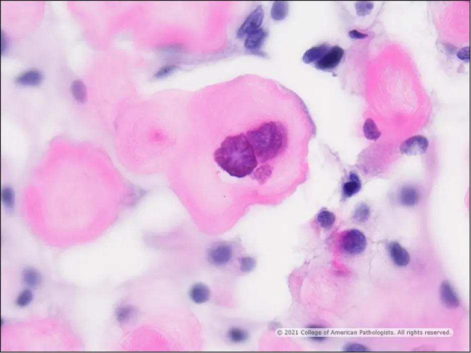
Image F: H&E. |
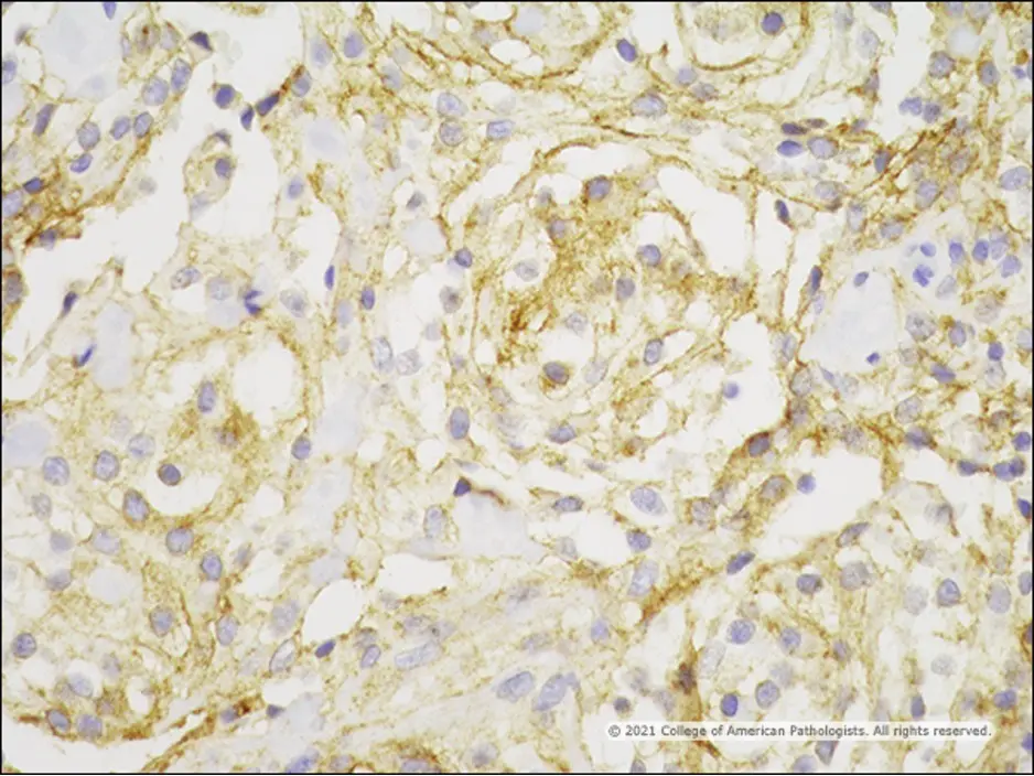
Image G: EMA. |
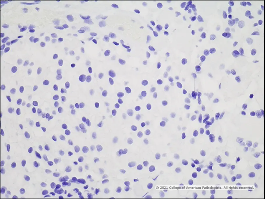
Image H: GFAP. |
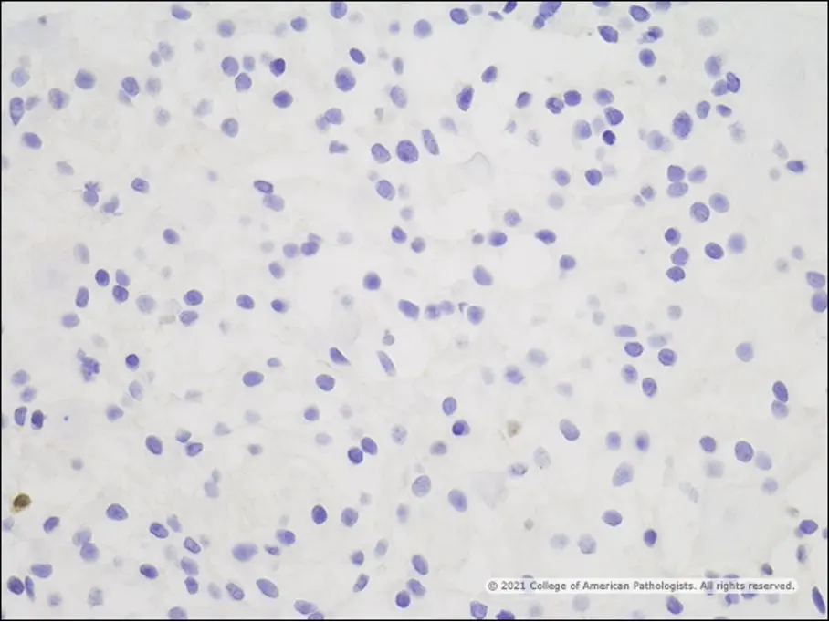
Image I: S100. |
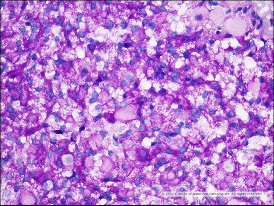
Image J: PAS. |
Familial cases of CCM have been associated with germline loss of function variants in SMARCE1. SMARCE1 (also known as BAF57) is a subunit of the SWI/SNF chromatin remodeling complex and functions as a tumor suppressor. SMARCE1 germline pathogenic variants predispose to both cranial and spinal CCMs.
The histologic differential diagnosis of CCM includes various tumors with clear cells including hemangioblastoma and metastatic clear cell renal cell carcinoma (RCC). Hemangioblastomas are highly vascular tumors with prominent thin-walled vessels. The neoplastic stromal cells of hemangioblastoma have finely vacuolated to foamy cytoplasm. Hemangioblastomas are immunoreactive for inhibin, D2-40, neuron-specific enolase, and S100 and are immunonegative for cytokeratin, EMA, and CD10. Vasculature can be highlighted with endothelial markers such as CD31 and CD34. Metastatic clear cell RCCs are composed of sheets of uniformly clear tumor cells that frequently have vesicular nuclei with prominent nucleoli. RCCs can also show other high-grade features such as hemorrhage, necrosis, and mitotic activity. RCCs are typically immunoreactive for cytokeratins, EMA, CD10, PAX-8, and RCC and are usually negative for inhibin.
The differential diagnosis of a cauda equina mass includes myxopapillary ependymoma and schwannoma, among other entities. Myxopapillary ependymomas are the most common intramedullary tumors in the cauda equina region. Histologically, they show a variably papillary architecture with tumor cells arranged around central hyalinized blood vessels with abundant Alcian blue-positive myxoid matrix in perivascular spaces. They can have variable numbers of microcysts filled with mucin. Tumor cells are immunoreactive for GFAP, S100, and occasionally EMA in a dot-like or surface staining pattern; they are negative for OLIG2. Schwannomas are composed of varying proportions of cellular Antoni A regions with compact spindled cells forming variable numbers of Verocay bodies and less cellular, loosely textured Antoni B regions. Degenerative nuclear atypia, hyalinized vessels, and hemosiderin deposition are common. Mucinous material may be present, particularly in the Antoni B regions. Tumor cells are diffusely and strongly immunoreactive for S100 and SOX10. EMA can be used to highlight perineurial-like cells within the tumor capsule. Collagen IV IHC or reticulin staining can demonstrate pericellular basal laminae.